Ark Vets have had a dental x-ray machine since 2011
Dental Radiography is an essential part of effective dentistry.
It provides us with 3 areas of information which are essential in the planning and carrying out of dental surgery:
- Dental Anatomy – there is a huge variation in the normal anatomy of teeth and their supporting structures (e.g. the skulls of a boxer and greyhound vary enormously). Teeth can have 1, 2 or 3 roots, but sometimes the number can vary or be fused. To allow us to effectively remove the teeth it is crucial for us to have this information
- Dental Pathology – it is essential in the assessment of disease within the tooth, and allows us to know which teeth are able to be saved and which need to be extracted
- Assessing Treatment – ensuring all affected teeth are removed completely.

Not just cats & dogs get their teeth x-rayed

Cat getting a dental x-ray taken
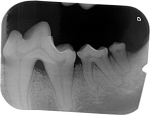
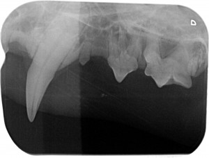
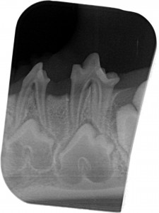
Some examples of dental x-rays
Shows both the adult and baby teeth in a dog

X-ray showing the jaw